Patient X-RAY Consent Form

Download NOW!

Have a Question?

info@readcts.com

Call Us

+1 (352) 505-7411

Our services

Login or Register

Login to access our platform or register if you're interested in our CT reading services.

Upload Your CT Scans

Please compress (Zip) your Dicom CT files before uploading them since this reduces the transmission time. For a guide showing how to log in and upload your files click here

Secure Technology

Patient radiographs, referral forms and reports are stored on an HIPAA compliant cloud server maintained by Citrix Inc. Data is backed up daily.

BENEFITS OF RADIOLOGIST REPORTS This can be summarized as better health care for your patients. Click to read more about this.
  • TIME It takes a specialist radiologist approximately 30 minutes to examine the whole volume of a CBCT and write a report. This is because each set of 512 axial, sagittal and coronal slices is read twice. First inferiorly for the anterior half of the volume from the paranasal sinuses to the lower border of the mandible and hyoid bone followed by superiorly from the cervical vertebrae, airway through the skull base and TMJs to the cranial vault. This is followed by coronally front of face to posterior skull and back again. Lastly sagittally from the right side of face to left and back again. Only after the whole volume has been examined for unknown pathology does the radiologist proceed to examine in detail the jaws for pathology. A panoramic reconstruction is made of just the maxilla and 0.1 mm thick slices examined for pathology. Any abnormalities are recorded as text in the report and if a picture will convey useful information to help the dentist manage the condition, a photograph is placed in the record. After this a new reformat of the mandible is created and the examination process is repeated. Why perform two separate examinations? Generally the radius of curvature for the maxilla is larger than the mandible so that if the maxillary teeth are in the focal trough then the mandibular teeth are not. This is particularly important for the third molars. Also the maxillary teeth often have an anterior proclination of the roots relative to the mandibular teeth. Unless you rotate the maxilla a little inferiorly to align the roots in the vertical position, you cannot see the whole root in a slice but only part of it. This is really important for tracing root canals and fractures. Additional time is needed to trace the Inferior Alveolar Nerve Canals and mark them in red. This is important for planning placement of implants. If anterior mandibular implants are planned, the location of the midline mandibular lingual foramen and blood vessel is marked by an arrow so that it is not cut by a drill outside of the bone. Although the lingual artery is a small vessel it can bleed for a long time into the sublingual space until the patient has difficulty in swallowing or breathing.

  • Systemic diseases Dentists who order a Panoramic or CBCT radiographic examination are responsible for reading the whole volume and detecting any diseases present. If they feel inadequately trained to do this, as most are regarding CBCTs, then they are responsible to have the images read by a suitably trained specialist to avoid failure to diagnose [Amer Acad Oral and Maxillofacial Radiology. Carter L et al Oral Surg Oral Med Oral Path Oral Radiolo Endod 2008 Oct;106(4):561-2. PMID 18928899]. How likely are you to find serious systemic disease? Calcification of atheromatous plaques in the walls of the external Carotid artery bifurcation and cavernous sinus portion of the internal Carotid artery are present in about 5% of adults over the age of 40 years. For people with high risk factors such as hypertension, obesity, hypercholesteremia or chronic kidney disease the prevalence rises to about 40%. In absolute terms a general dentist during their practicing life time could be expected to see hundreds of their patients who have detectable carotid artery calcifications in both panoramic and CBCT images. The correct management is to inform the patient that a calcification has been detected which could indicate that they have cardiovascular disease. They should consult an MD to see if further investigations are required. For those patients who are not aware that they have cardiovascular disease this referral could save their lives. To place this in perspective, although oral cancer screening is important, a dentist in their practicing lifetime will probably only diagnose 3 - 10 cases.

  • Radiology reports The radiology report is divided into two main sections: a) Single page of text, b) Images - a panoramic image and up to six additional images. The text page is divided into Dental Findings, Specific Findings, TMJs, Sinuses, Airway, Cervical spine, Calcifications and finally Impressions where a summary of the findings are presented with indications of the severity, need for further tests, monitoring intervals and referral if appropriate.

    Sample Reports: 
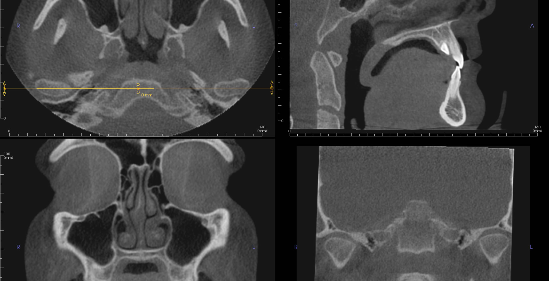
Carotid Artery Calcifications 
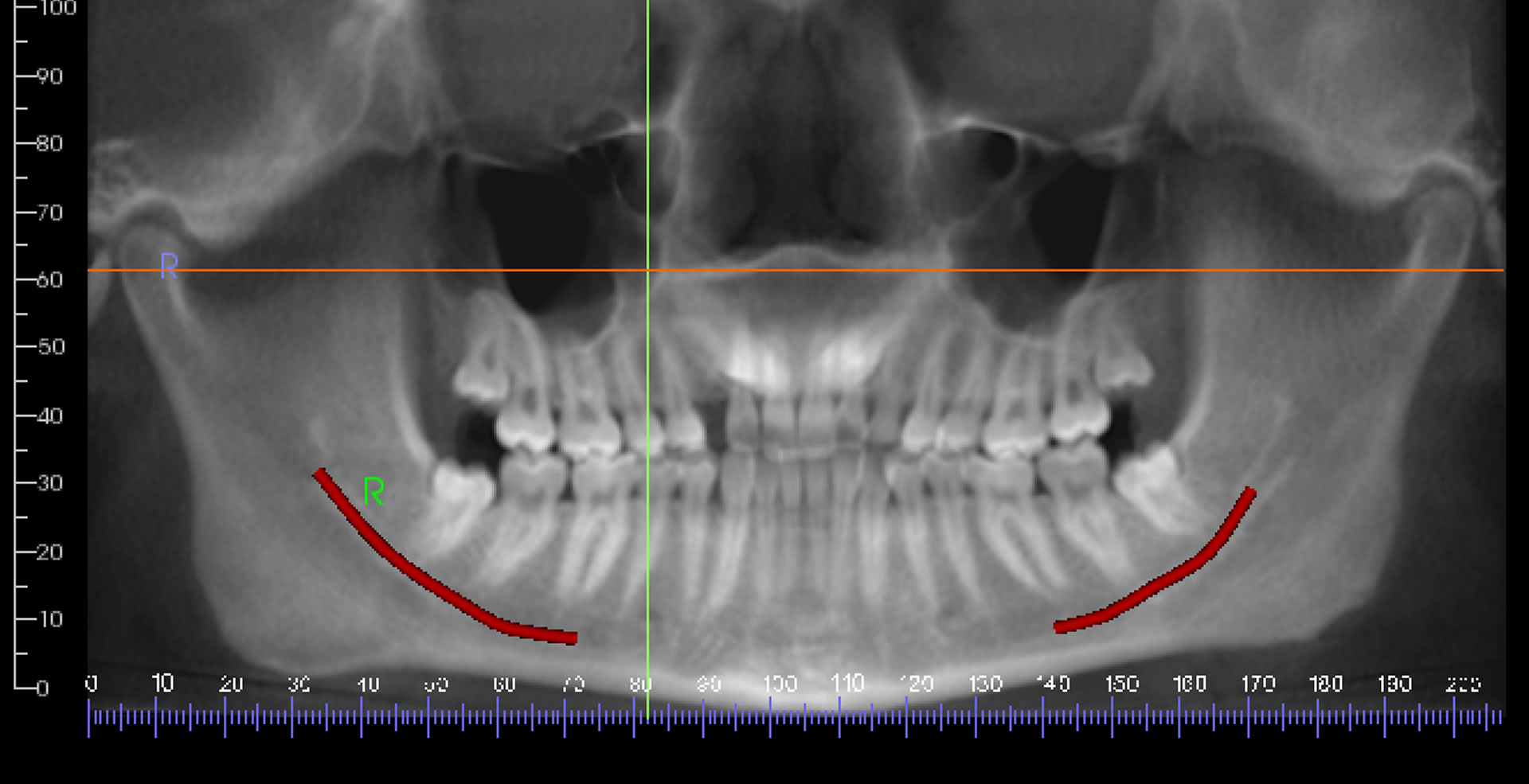
Pre-Implant Assesment
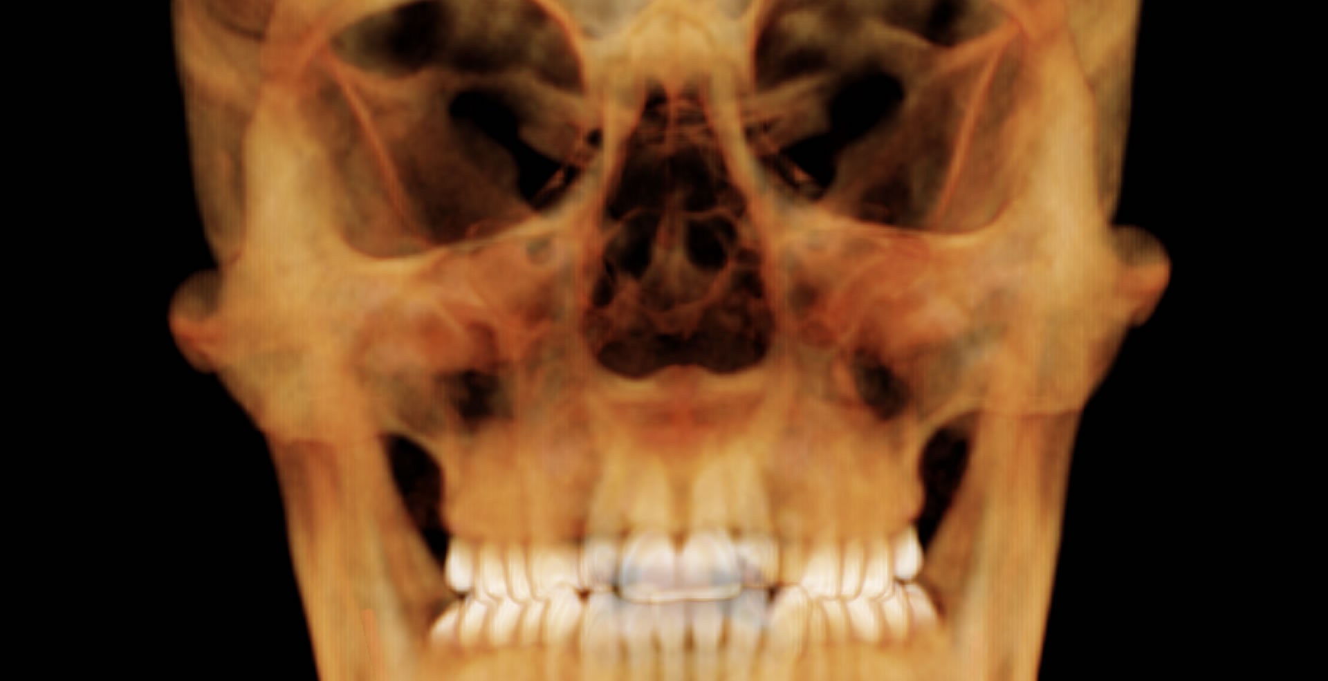
 Third Molars 
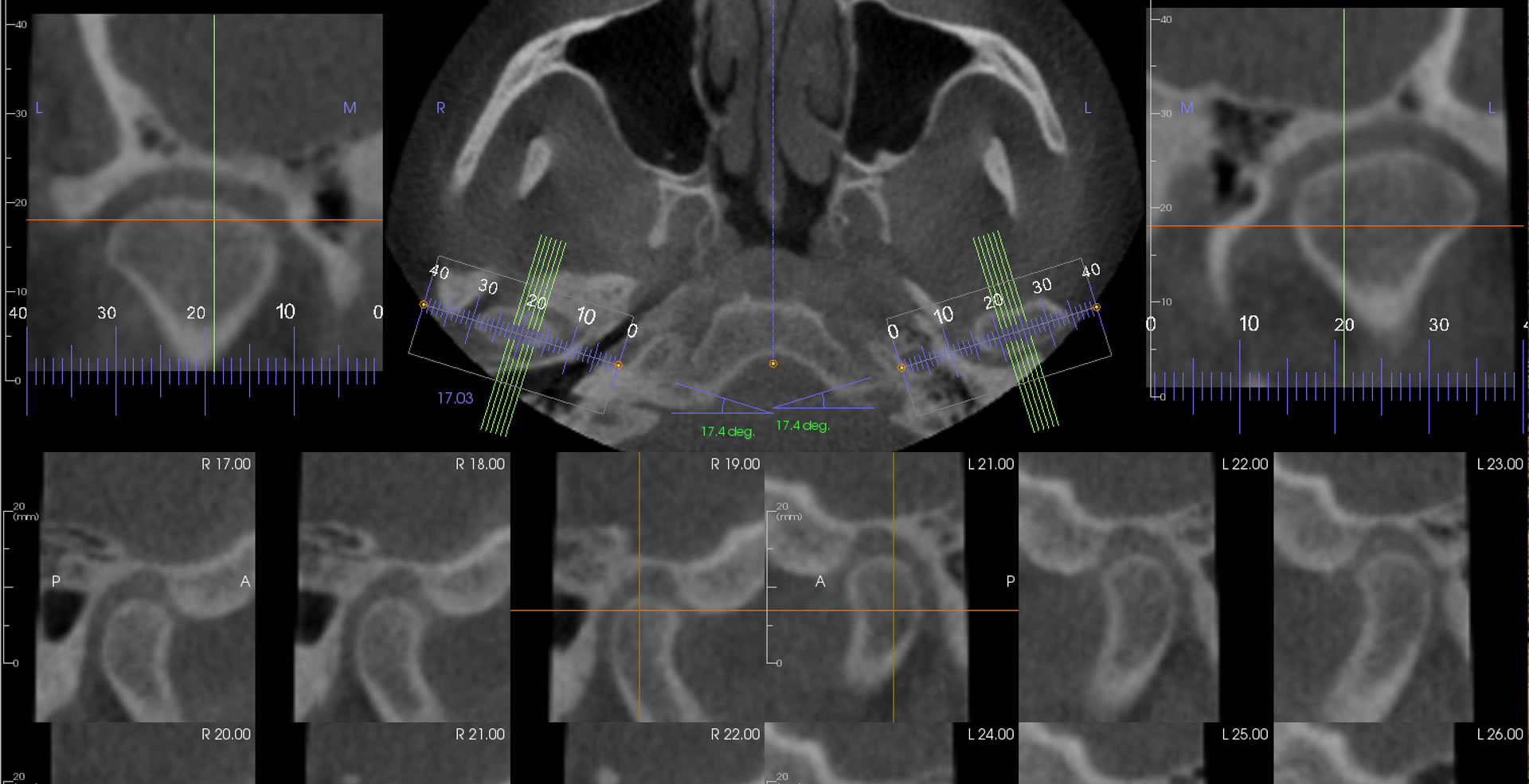
Temporomandibular Joints

  • Dentist / Radiologist collaboration Just as you develop a close relationship with your dental laboratory to create the best results for your patients, the same is true with radiologists. Reports can always be tailored to your special needs be it endodontics, prosthodontics or any other area. TMJ examinations can be useful for showing bone pathology but CBCT is of little value for TMD patients where the pain is originating in muscles, ligaments or cartilagenous discs since these are not visible in X-ray radiographs. However, visualization of boney changes due to degenerative joint disease or rheumatoid arthritis is helpful in managing TMJ disease.

About Us

50

Year of Experience

28

Years of Teaching and Reporting Radiology

10

Or more Patents

50

Or more Refereed Scientific Publications

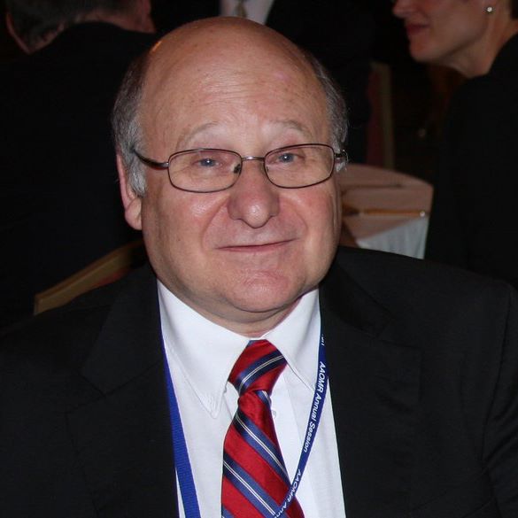
Douglas K Benn BDS, DDS, PhD, trained as an Oral and Maxillofacial radiologist at Guy's Hospital, University of London and the Royal College of Radiologists, England. For 12 years he was tenured Full Professor of Maxillofacial Radiology at the University of Florida College of Dentistry and Full Professor of Radiology in the Department of Diagnostic Sciences, Creighton University School of Dentistry, Omaha, Nebraska.
Dr. Benn is an author of more than 50 refereed scientific papers on radiology and the inventor of several patents. He was Editor-in-Chief of Dentomaxillofacial Radiology, the peer reviewed journal of the International Association of Dental and Maxillofacial Radiology.

Why Choose Us

Reports in English and Japanese
Secure Upload Service
Online reports

FAQ

Is your Upload Service secure ?

https://www.ReadCTs.com is the Home site of Dental and Maxillofacial Radiology Omaha LLC and is a secure website as indicated by the . Patient referral information collected by the Referral form is stored on a secure HIPAA compliant server hosted by Citrix Inc.
Upon completion of the patient referral the user is automatically redirected to login to the https://www.ReadCTs.sharefile.com site hosted by Citrix where they can upload their CBCT Dicom files into their folders. Dental offices can only access their own folders and no others. When a report is ready to download access is only granted to a restricted set of folders.

How do I log in to your Upload Service ?

Click here for full instructions to Register as a New Office, Login, complete a Referral Form, download reports.

Can I have a Sample Report ?

How much is the payment for your service ?

Fees:
CBCTs - Single site/single jaw US$ 97.00
Double jaw US$ 112.00.
NOTES: Full volume is examined for any pathology.
Implant cases Routine tracing of Inferior Alveolar Nerve Canal (IANC) and measurement of edentulous alveolar bone ridge height and width.
Third molar removal Position of tooth apices relative to the IANC and maxillary sinus floor, evaluation of tooth follicle size, root resorption of adjacent teeth and loss of periodontal attachment due to impaction.
TMJs Charged as a double jaw since all teeth and jaws in volume have to be examined.
Cleft palate assessment Charged as a double jaw due to the complexity and time needed for assessment.
Root canal therapy Single jaw or double. Routine examination of number of canals present unfilled or filled, location of filled root canal so if eccentric location in upper or lower molars then possibility of missed MB2 or additional canals. Furcation bone loss but no apical radiolucencies then possibility of root fracture. Mid-root radiolucency without apical pathology then possible fracture or root stripping from endodontic treatment. Consider periodontal disease, neoplasia, Bisphosphonate-related bone necrosis. Under- or over-extended root fillings.
Panoramic Radiographs - US$ 50.00.
Intra-oral radiographs - US$ 30.00.

Contact Us